Solid State Quantum Computation
with Spins in Silicon
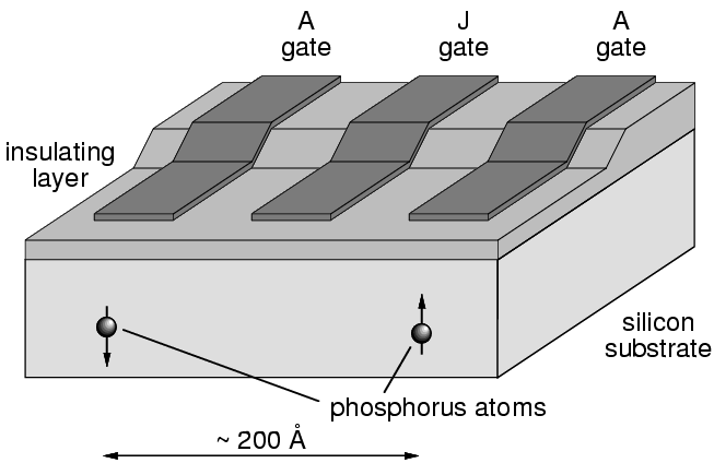
Quantum computation is a revolutionary new computational paradigm with compelling real-world applications that would provide computational capabilities far exceeding those of any existing or future conventional computer. Solid state implementations appear most likely to be able to provide a scaleable architecture and the potential for large scale fabrication, and spin degrees of freedom (both nuclear and electronic) embedded in such solid state systems are very attractive candidates for quantum bits (qubits). Kane has recently published a break-through conceptual study for a silicon based quantum computer based on nuclear spins that meets these requirements [B.E. Kane, Nature 393, 133 (1998)]. Other solid state schemes based on electronic spins have also been proposed. A common feature of these schemes is the requirement for single electron spin readout.
The magnetic resonance force microscope (MRFM) is a novel scanned probe instrument which combines the three-dimensional imaging capabilities of magnetic resonance imaging (MRI) with the high sensitivity and resolution of atomic force microscopy. Rapid advances in force detection sensitivity make mechanical detection of magnetic resonance dramatically more sensitive than conventional inductive detection; so much so that single electron spin detection appears achievable. The MRFM is thus excellently suited to meet the device characterization and qubit readout requirements of a spin-based quantum computer. We propose to optimize the sensitivity of the MRFM with the objective of detecting the magnetic resonance signal from an individual electronic moment. Combining single spin detection and controlled sub-surface sensitivity, the MRFM will meet two crucial needs for the silicon based quantum computer: spin qubit readout and device characterization.
The Silicon Based Quantum Computer
Single Spin Readout with the MRFM
Perhaps even more challenging is the single spin qubit state readout required at the end of the calculation. The hyperfine coupling between the nuclear spin qubit and its associated electronic moment allows qubit readout to be accomplished by determining the electronic spin state, so the ability to detect a single electronic moment will be the crucial tool for meeting this challenge. The single spin MRFM would be well suited to this task: it directly measures the spin state of an individual electronic moment, and the magnetic resonance imaging capabilities stemming from the presence of the magnetic field gradient give the MRFM the ability to select the individual electronic moment to be queried. The ability to select individual spins means that in conjunction with rf pulses to control the nuclear spin, this technique can also be used for qubit initialization.
In our laboratory we are operating the scanning Magnetic Resonance Force Microscope that operates down to 4 K and has the magnetic probe tip incorporated into a force detector which can be scanned over the sample using a conventional piezotube scanner. This unique instrument provides high spin sensitivity in fully scanning microscope. The microscope also functions as a scanning AFM to provide crucial sample topography information as a prelude to magnetic resonance scans. Presently our force detectors have a sensitivity of approximately 50 atto Newton (aN) in a 1 Hz bandwidth, (1 aN =10-18 N) and the field gradients generated by the miniature magnets (with sub-micron tip radii) we deposit on the cantilever tip are ~104 T/m. Optical displacement detection adds no significant noise in comparison to the displacement noise of the cantilever. Hence this microscope has a spin sensitivity of approximately 103 fully polarized electronic moments.
UHV 3He Cryostat for Ultrahigh Sensitivity
Temperature is one of the most important parameters determining microscope sensitivity. We will use 3He refrigeration, capable of cooling to 250-300 mK to cool our microscope and achieve high sensitivity. Though the lower temperatures achievable with dilution refrigeration might make that seem to be the best approach, we find that 3He refrigeration with its higher cooling power and its capacity for providing a UHV environment at room temperature provides the best approach to single spin MRFM.
The generation of MRFM signals requires manipulation of the sample spin magnetization by means of radio-frequency (rf) or microwave frequency oscillating magnetic fields which irradiate the entire sample and the sample surroundings. These fields can generate substantial unwanted heating. The large cooling power of a 3He refrigerator enables it to handle these large heat loads.
The problem of dissipative probe-sample interactions also argues for 3He refrigeration. A microscope allowing UHV surface preparation and characterization prior to sample transfer to the ultra-low temperature MRFM without breaking vacuum. This will allow studies of these dissipative interactions so that they can be eliminated or minimized. UHV can be achieved in a 3He refrigerator, and this is the route we are presently pursuing in our laboratory.

R. G. Clark, Director of the Centre for Quantum Computer Technology with nodes at the University of New South Wales, the University of Queensland and the University of Melbourne in Australia.
