|
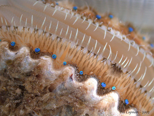
| Scallop eyes |
The Human Visual System
The structure of the eye
The iris and adaptation
Most of the eye's adaptation to dark and light occurs in the retina.
This adaptation is chemical in nature.
The retina primes more or fewer photo-receptor molcules to receive light
depending on the ambient brightness.
For young eyes, it takes 15 to 20 minutes for the retina to adapt to changes
in brightness.
As one ages, the adaptation time lengthens to nearly an hour.
Under any one circumstance of lighting, the retina has a dynamic range of
about 1000 to 1.
That means one can see detail in surfaces differing by a factor of 1000
in brightness.
Brighter surfaces will be washed out and hard to look at, while dimmer surfaces
will appear black with no detail.
Including adaptation, the dynamic range becomes a million times more to
one billion to one.
For comparison, photographic film has a dynamic range of about 100 to 1 and
CCDs 5000 to 1.
Aberrations and near / farsightedness
Near-sighted (myopic) eyes have too much lens curvature (the focal length
is too short) to be able to focus images of distant objects.
Myopic vision requires negative curvature (thin in the middle) lenses
for correction to reduce the overall curvature of the system.
Far-sighted (hyperopic) eyes have too little lens curvature
(the focal length is too long) to be able to focus images of near objects.
Hyperopic vision requires positive curvature lenses for correction.
As one ages, the flexibility of the lens decreases, tending toward
far-sightedness.
The image contains large field distortions since it is focused on a spherical
retina, but these distortions are easily corrected in the brain in a
process called constancy.
Scattering
The choroid and red-eye reflection
Some cameras have a pre-flash that momentarily reduces the subject's pupil size and
so reduces the amount of light entering the eye and the amount coming back through
the lens.
Nocturnal animals have a special coating on the choroid called the
tapetum lucidum.
The tapetum lucidum serves to reflect light back through the retina, increasing the
quantity of light caught by the photo-receptor cells.
It is this reflection one sees from animals in car headlights at night.
Different species can be identified by the color reflected from their eyes.
The retina and the brain
The visual field and the
lateral geniculate nuclei
The left fields of each eye are processed in the right LGN, and the right
fields are processed in the left LGN.
The cross-routing of the axons occurs in the optic chiasma.
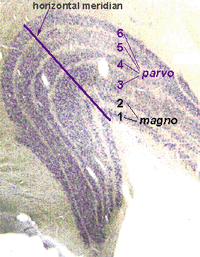
The image (still latent) is more-or-less preserved in the LGNs.
LGN cells are arranged in layers, with cell neighbors in one layer
corresponding to ganglion neighbors in the retina.
Corresponding image locations are stacked "above" each other in
adjacent layers.
Color information goes through layers 3-6, while bright-dark information
goes through layers 1 and 2.
In the cross section above, the line marked horizontal meridian separates
the processing of the upper visual field (below the line) from the processing
of the lower visual field.
Comparison of the layers (one from one eye and another from the other
eye) is the first step toward
binocular depth perception.
Information processed in the LGNs continues on to the
visual cortex.
These modules will not consider what happens in the cortex.
Summary:
The lens modifies the image focus by adjusting its focal length, called
accommodation. Near-sighted (myopic) eyes focus the image in
front of the retina. Far-sighted (hyperopic) eyes focus the image behind
the retina.
The sharpest part of the image is focused on the fovea of the retina
(on axis behind the lens).
The retina contains five layers of cells and cell connections.
The photosensitive cells (rods and cones) are at the back.
The other layers
perform complex interconnections before accumulating the signal in the
ganglion cells.
The ganglion cells have long axons (signal carrying threads) leading
accross the retina, through the optic nerve bundle,
to the lateral geniculate nucleus in the brain.
The field of view of each eye is split in half. The left fields are
processed in the right LGN, and the right fields are processed in the
left LGN.
The cross-routing of the axons occurs in the optic chiasma.
Most of the eye's light-dark adaptation is chemical. The sensitivity
of the photo-receptors depends on the amount of light falling on them.
More light decreases the sensitivity.
The iris acts almost instantaneously to help protect the retina from
overstimulation, but it can change the light level on the retina
by only a factor of 10 or so.
Some comments on the evolution of the eye.
Sample questions for reflection
Be able to recognize the correct order of eye parts encountered by
incoming photons.
How does the eye focus an image on the retina?
What type of lens (positive = thick in the center, or negative = thin in the
center) is required to correct near-sightedness? far-sightedness?
How does the eye manage aberrations?
What wavelengths is the eye least able to focus?
What are the photosensitive cells on the retina?
Where are the photosensitive cells in the retinal layering?
What is the fovea?
What processes are used to adapt to different levels of lighting?
Which cells pass signals from the retina to the LGN?
There are two LGNs, one in each side of the brain. What is each
responsible for?
What is meant by the term visual field?
Be able to recognize the correct paths of ganglion axons from the
eyes through the chiasma to the LGNs.
How are LGN cells arranged?
Where do visual signals go after the LGNs?
The eye is roughly spherical, with the cornea and lens in front, and the retina
on the back inner surface.
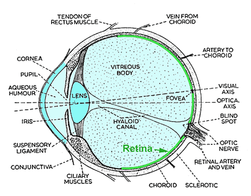
Most of the refraction required for focusing an image on the
retina occurs at the air-cornea interface.
The lens modifies the image focus by adjusting its focal length.
This process is called accommodation.
The ciliary muscles pull the lens into the proper shape.
This webpage has an applet demonstrating accommodation.
The sharpest part of the image is focused on the fovea of the retina
(on the visual axis behind the lens).
The iris (Greek rainbow) controls the aperture, or pupil, of the
eye.
In strong light, it closes down to help protect the retina from overexposure.
In darkness, it opens to admit as much light as possible.
The iris reacts almost immediately to changes in light.
You can test your iris reaction time by going into a dark room with a mirror,
and turning on the lights while looking at the mirror.
Most aberrations
in the cornea-lens system are effectively minimized
by the non-uniform index of refraction in the lens.
Some chromatic aberration remains.
It is easiest to experience this aberration at night with distant blue
neon signs.
Short waves are focused too close to the lens for the eye to accommodate the
image on the retina.
In the daytime, the eye aperture (pupil) is smaller so the depth of field
is larger.
In addition, there is nothing important in nature that is bright blue-violet,
so our eyes have not evolved to choose this wavelength range for focusing.
As light passes through the cornea-lens and humor fluids in the eye, it has
many opportunities to scatter off edges of the iris and defects in the materials.
Normally one does not notice this scattering in the daytime, because the background
image is bright and the brain removes variations induced by the eye via constancy.
However, at night when the background is dark, the scattering becomes part of the
image and is noticed.
Examples include the rays seeming to spread out from street lights and/or on-coming
headlights, and the "starriness" of stars.
The choroid is the back support of the retina and contains layers of blood vessels
that nourish the back of the eye.
When a camera flash goes off, the light enters the eye and is focused on the retina
as a very bright spot.
The cornea-lens re-focuses light scattered from that spot into a nearly parallel
beam heading back toward the camera (remember light paths are always reversible).
The scattered light is red from blood.

Stop-and-Go kitty
The retina contains five layers of cells and cell connections.
The photo-sensitive cells (rods and cones) are at the back.
The other layers perform complex interconnections to accumulate the signal into
ganglion cells.
The ganglion cells have long axons (signal carrying threads) leading
accross the retina, through the optic nerve bundle,
and ending in the lateral geniculate nuclei in the brain.
Some scientists think of the retina as a part of the brain, since it
is intimately connected and performs complex analysis within its own structure.
The visual field is the part of the world imaged on the retina of each eye.
With the left eye one can see from almost 90° to the left to about 45°
to the right of straight ahead.
The right eye field is similar but shifted right.
The visual fields overlap for about 90° in the middle.
The field of view of each eye is split by a vertical line through the fovea of
each retina.
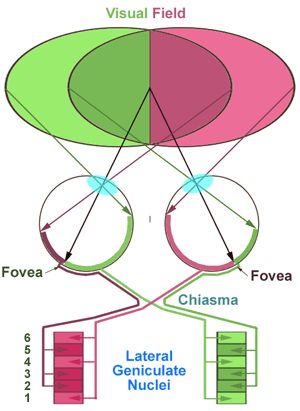
The path from the visual field
to the lateral geniculate nuclei.Cross section of a
lateral geniculate nucleus
The eye is roughly spherical, with the cornea and lens in front, and the retina
in the back. Most of the refraction required for focusing an image on the
retina occurs at the air-cornea interface. The iris acts as an aperture
control mechanism.


What is meant by the term accommodation?
Keep in mind the difference in optical meaning between adaptation and
accommodation.